Introduction: In the medical field, performing those complex surgeries related to the brain or skull can be a very difficult task even under the best medical conditions. However, the difficulty of surgery increases exponentially when it involves infants and children, especially newborns.
On August 13, 2022, in a recent surgical case abroad, doctors used3D printing technology to successfully help save a newborn baby with a similar condition. The newborn reportedly needed surgery immediately after birth to repair an occipital defect in his skull, as it had not developed properly. To address this challenge, Polish deep tech company Sygnis SA (which recently acquired Zmorph) used additive manufacturing technology to create a 3D printed model that was critical to the success of the newborn baby's neurosurgery.

In addition to being used in prosthetic and orthotic manufacturing, another major way 3D printing is being used in the medical field is for training and preparation prior to surgical procedures. Doctors can do this by printing an exact replica of the area to be surgically treated, in the case of this article, a model of the patient's skull made from a scan of the patient. Surgeons are able to practice before surgery, thereby reducing mortality, especially for complex procedures like this one. To help save this particular baby girl, Pawel Ozga, a medical image segmentation specialist and volunteer with the Polish e-Nable Foundation led by Dr. Kryzstof Grandys, approached Sygnis to create the model as soon as possible and in as much detail as possible to increase the likelihood of survival. This child was diagnosed with an occipital defect immediately after birth, which also meant that part of her brain was exposed, a dangerous condition that can be fatal if left untreated.

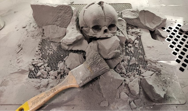
△ SLA model of a newborn's skull, including the defect that needs to be repaired (Image credit: Sygnis SA)
Creating a 3D printed skull model
The creation of the pre-surgical skull model was performed in February 2022. After the baby was born, she was diagnosed at the University Children's Hospital in Krakow. the 3D printed skull was a key part of the pre-surgical plan as a way to practice with an accurate model of the baby's skull in a short period of time. In fact, Sygnis actually created two models, one using SLA and the other using SLS. they turned to these two different technologies to take advantage of the benefits of both.
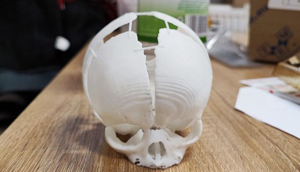
For the model created using SLA, it was done on a SygnisFlashforge 8.9s 3D printer. the benefit of SLA is that the doctor is able to get a more detailed and accurate reproduction of the skull based on MRI and CT scans. However, the material used is not as strong. That's where SLS comes in, this model was created on a Sinterit Lisa Pro 3D printer. SLS also produces highly detailed and accurate parts, but they are much stronger due to the use of PA12. This means that doctors are able to actually practice with their tools before surgery. In addition, because SLS 3D printing does not require a support structure, but rather a powder blank to fill this role, doctors were able to perfectly reproduce geometrically complex skeletal structures. By being able to practice with both models beforehand, the doctors were able to prepare quickly and efficiently for this difficult procedure.
In addition to the ability to perfectly reproduce the baby girl's skull from the scan alone, the doctors chose 3D printing technology because of its ability to create this 1:1 scale 3D printed model of the skull at an efficient manufacturing speed. Since the defect was not diagnosed during the pregnancy, time was of the essence to complete the procedure in the shortest possible time. In fact, according to Sygnis' case study, they only had 96 hours to create the model, deliver it, and have the surgeon perform pre-surgical testing. Thankfully, the skull printed using SLA was completed in just eight hours, while the skull made using SLS was completed in 24 hours.

△ By using both SLA and SLS (pictured) models, the surgeon was able to practice effectively before the surgery, despite the time constraints (Photo credit: Sygnis SA)
The surgery was successfully completed on February 28 and the little girl was able to be discharged from the hospital. Professor Lukasz Krakowczyk, the surgeon in charge of the operation, concluded: "Imaging studies are helpful in identifying cranial defects. However, they don't exactly match the skin defects, which is why printing the model is so important. 3D printing will also be essential at the stage of reconstructing the cranial defects, when the bone reconstruction needs to be perfectly aligned and planned."
Although the final outcome cannot be known, as this child will need more treatment in the future, it is clear that this surgery was able to be successful thanks to the intervention of 3D printing. sygnis also expects more such collaborations, as they further use hand preoperative models to improve the success rate after surgery.


